


TL;DR
Kuffler’s 1953 paper is a foundational text in visual neuroscience, introducing the concept of the "center-surround" receptive field. By recording from single cat retinal ganglion cells, Kuffler demonstrated that these neurons are not simple light detectors but spatial filters that use antagonistic excitatory and inhibitory zones to enhance contrast and detect edges.
What Problem Does This Paper Address?
Before Kuffler, the retina was often viewed as a relatively simple relay. While researchers like H.K. Hartline had identified that some optic nerve fibers responded to light onset ("on") and others to offset ("off") in frogs, the spatial logic of these responses in mammals was unknown. Kuffler aimed to map the "receptive field"—the specific area of the retina that influences a single ganglion cell’s firing—to understand how the retina organizes visual information before it ever reaches the brain.
Methods
Kuffler’s experimental setup was a feat of 1950s precision engineering. He developed a "Multibeam Ophthalmoscope" that allowed him to project tiny, independent spots of light (as small as 0.1 mm) onto the retina of an intact cat eye while simultaneously recording from single ganglion cells.
Key technical aspects included:
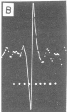
- Microelectrode Recording: He used fine platinum-iridium wires (10-15 µm) to record extracellular potentials. He learned to distinguish between signals from axons (passing fibers) and the ganglion cell bodies themselves by analyzing the spike's polyphasic shape (Fig 1).
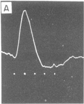
- Pressure Blocking: To prove he was recording from a single unit, he used a "pressure block" technique (Fig 2). By applying slight pressure with the electrode, he could cause the main spike to fail, revealing a "prepotential"—the local synaptic input driving the cell.
- Controlled Adaptation: A third light source provided adjustable background illumination, allowing him to study how the retina's behavior changed between light-adapted (photopic) and dark-adapted (scotopic) states.
Datasets
Not discussed in the paper (this research predates modern digital datasets; data consisted of electrophysiological recordings from individual neurons in cats).
Key Insights and Ideas
- The Concentric Map: The most famous finding is that receptive fields are organized into concentric circles (Fig 6). A cell might have an "on" center and an "off" surround, or vice versa.
- Spatial Antagonism: The center and surround are not just different; they are antagonistic. Light hitting the surround inhibits the response generated in the center. This "push-pull" architecture means the retina is tuned to detect spatial gradients (edges and contrast) rather than absolute, uniform brightness.
- Dynamic Receptive Fields: The size and sensitivity of these fields are not fixed. They "shrink" under bright background light and expand in the dark. This suggests the retina's spatial resolution is dynamically linked to its adaptation state.
- High Bandwidth Signaling: Kuffler recorded firing rates as high as 800 impulses per second—near the physiological limit for neurons—with latencies as short as 15 ms, highlighting the high-speed nature of visual processing.
Key Results
- Response Flipping: By shifting a 0.2 mm light spot across the retina, Kuffler showed he could transform a cell's response from a pure "on" burst to a pure "off" burst just by moving the stimulus 0.5 mm (Fig 4).
- Field Dimensions: Receptive fields in the cat retina were found to span roughly 1-2 mm in diameter.
- Interaction Effects: He demonstrated that the "on-off" response seen with large-field illumination is actually the result of the summation and interaction of antagonistic center and surround signals.
Limitations and Future Work
- Light Scattering: The optical imperfections of the eye caused light to scatter, making it difficult to define the absolute, sharp boundaries of the receptive fields.
- Sampling Bias: The microelectrode technique likely favored larger ganglion cells, potentially missing the behavior of smaller, more numerous units.
- Anesthesia: Kuffler noted that anesthetics like Dial-urethane suppressed spontaneous activity, which might alter the "natural" state of retinal signaling.
- Future Directions: The author suggested that similar antagonistic organizations might exist in higher brain centers and that studying animals with foveas (like monkeys) would be the next step to understanding human vision.
Summary updated